


Classifying disease evolution
DISEASE STAGE
The stage of a tumor reveals its development; it reflects how deeply spread it is in the intestinal wall, and if it has dispersed to the lymph node or to other tissues or organs.
The classification is done using clinical reports, blood tests, imaging exams (TACs, MRIs, X-rays, PET, etc.) and can be further updated after surgery (e.g., with information from the tumor biopsy and the extracted lymph nodes).
The stages here presented are a simplified version of a complex analysis called TNM (that analyzes T=tumor, N=nodules, M=metastases).
Higher disease grades correspond to bigger and/or more disseminated tumors.
Further to TNM, there are other ways to measure tumor evolution/stage.
One is called the Dukes' staging system.
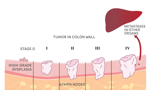
STAGE 0
The cancer remains at its original point (in situ, Latin for in position) inside the colon and/or rectum. Carcinoma-in-situ is the other name for colorectal cancer stage 0
STAGE I
The tumor has grown inside the colon and rectal wall. For now, however, it has not reached their exterior wall, or pass to the outside of the colon
equivalent: Duke’s A
STAGE II
The tumor has grown deeper into the wall of the colon or even through it. It may have invaded the surrounding tissues, but the cancer cells have yet not reached the lymph nodes.
equivalent: Dukes’ B
STAGE III
The cancer is larger and may have spread to the surrounding tissues and/or the lymph nodes
equivalent: Dukes' C
STAGE IV
The tumor has spread (metastasized) to other parts of the body, such as the liver or the lungs.
equivalent: Dukes' D